How to view anatomical structures. A 2-month-old girl presented with a lesion on her tongue that had evolved over 2 weeks.

Heart Anatomy Labelled Illustration Stock Image C043 4821 Science Photo Library
Surgical resection of the lesion was performed.
. It primarily functions as a lymphoid tissue although it has an important endocrine function that involves the production of thymosins. B Magnified schematic of AIB and its neighborhoods in A C Representative confocal image showing the lateral view of an AIB neuron labeled with cytoplasmic mCherry cyan. Two granular areas referred to as Ig1 and Ig2 insular lobe granular areas found in the dorsal posterior insula and a dysgranular area labeled Id1 d for dysgranular area found in the.
Materials Microscope Slides LABPAQ Kit. We will view the histology during the lymphatic and immunity unit. Mitral valve labeled at center right Base of ventricles exposed by removal of the atria.
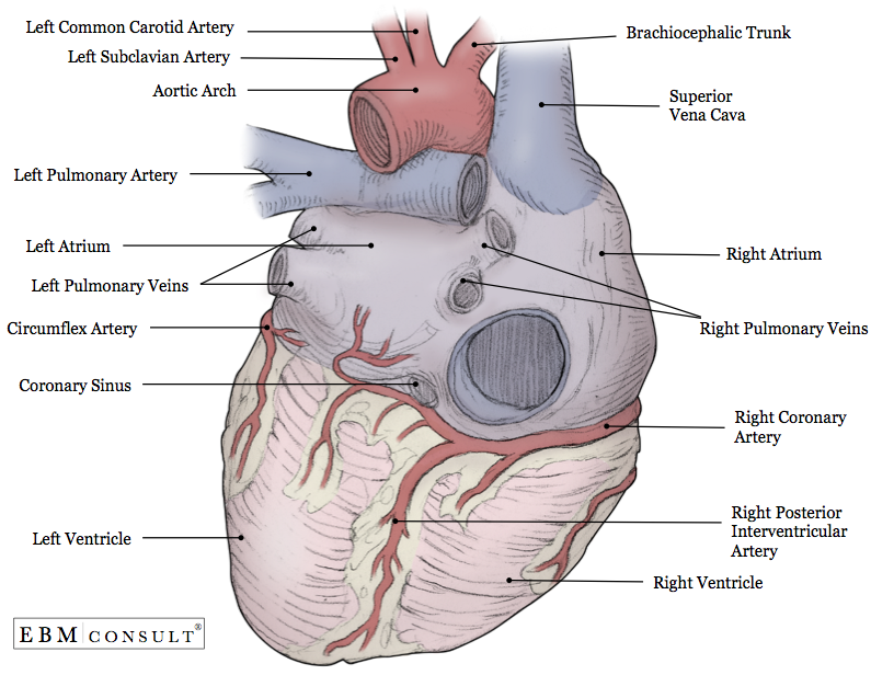
Midway is the central compartment of the thoracic cavitySurrounded by loose connective tissue it is an undelineated region that contains a group of structures within the thorax namely the heart and its vessels the esophagus the trachea the phrenic and cardiac nerves the thoracic duct the thymus and the lymph nodes. Histopathologic study showed a. A Anterior view of the external heart C 2019 Pearson Education.
This tool provides access to an MDCT atlas in the 4 usual planes allowing the user to interactively discover the heart anatomy. The mediastinum from Medieval Latin. Aortc arch Ligamentum arteriosum Left pulmonary artery Left pulmonary ve ns Auricle of left atrium Circumflex artery Left coronary artery in atrioventricular sulcus Great cardiac vein Left ventricle Anterior interventricular artery in anterior interventricular sulcus Apex.
Tricuspid valve visible at bottom right Details. Bicuspid mitral valve visible at bottom left. The quiz mode makes it possible to evaluate the users progress.
The images are labeled providing an important medical and anatomical tool. The physical examination revealed a tumor hard in consistency located in the posterior midline of the tongue with a base of approximately 1 cm and 2 cm in length. The only study to comprehensively analyze the cytoarchitecture of the human posterior insula using an observer-independent approach points to the existence of three distinct areas therein.
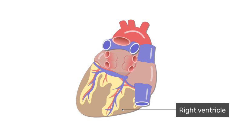
Anterior frontal view of the opened heart. White arrows indicate normal blood flow. Valva atrioventricularis sinistra valva mitralis valvula bicuspidalis.
The thymus is located anterior to the heart distal to the thyroid. Anatomy of the human heart and coronaries. Unspecified systolic congestive heart failure I5021 Acute systolic congestive heart failure I5022 Chronic systolic congestive heart failure I5023 Acute on chronic systolic congestive heart failure I5041.
Anatomy Heart External

Posterior View Of The Heart Heart Anatomy Heart Diagram Anatomy

Heart Posterior View Diagram Quizlet

Posterior View Of The External Heart Diagram Quizlet
The Heart Chambers And Their Functions

Posterior View Of The Heart Diagram Quizlet

4 Posterior View Of The Human Heart Download Scientific Diagram

Heart Anatomy Anatomy And Physiology Ii
0 comments
Post a Comment